Showing posts with label brain. Show all posts

Monday, October 29, 2012

Medical Monday: Down syndrome, Alzheimer Disease and a Very Special Mouse

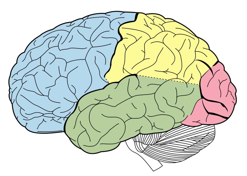
In the last two posts for Medical Monday I have outlined the basic anatomy of the brain and have given an overview of some of the differences between a typical brain and one with Down syndrome.  Today's post is about some of the research that is going on that concerns Down syndrome and why that is significant. 
One of the major areas affected by Down syndrome is the hippocampus, which is largely tasked with spacial skills and memory creation/consolidation.   As there are three copies of chromosome 21 with Down syndrome (Trisomy 21), one theory is that it is an over expression of specific genes which causes some of the major cognitive difficulties. 

If the specific genes themselves, cellular activity created by these genes or even a protein or compound secreted/lacking from the cells can be targeted, some of the cognitive problems with DS can be alleviated (or so the theory goes). Most of the study in this area however,  could not be accomplished without a very special mouse.


The Down syndrome Mouse Model

Photo courtesy of Stanford School of
Medicine, Down Syndrome Research Center
Most of today's research involves a type of mouse that has been bred to have many of the characteristics of DS that occurs in humans.  We share many genes with mice;  the mouse chromosome 16 contains 80% of the genes of the human chromosome 21 (including most of what is known as the DSCR or Down syndrome Critical Region).  Mice with a trisomy of chromosome 16 have been available for decades, however none survived very long.  With the creation of new mouse models that do survive, there is an ideal analog that reproduces fast and readily.  These mice share many features with human DS:  developmental delay, cranio-facial changes, learning issues, hyperactivity, weight problems and some behaviour issues.  However, it is not a perfect model as it does not cover all of the DSCR;  new mouse models will need to be developed in time.

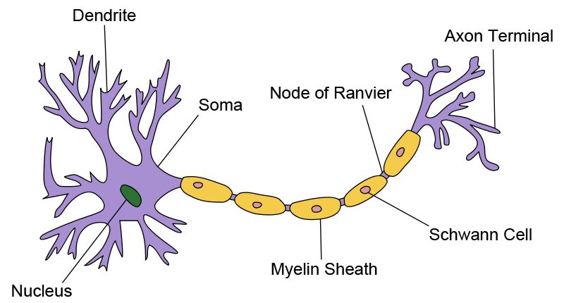
Having a model to help understand things like cognition has proved to be infinitely helpful and has opened many new avenues of research into Down syndrome, cognition and aging.   Included are: the excitation/inhibition of certain neural pathways, restoring neuronal pathways, neurotransmitter restoration, sleep abnormalities and the discovery/reassignment of drugs for treatment (for more information regarding specific American projects, see this listing, courtesy of the Down Syndrome Research and Treatment Foundation.  For a Canadian listing, see the Down Syndrome Research Foundation).


Down Syndrome and Alzheimer Disease

Alzheimer Disease is the most common form of dementia and has a very early onset in the Down syndrome community.  As a result, these two areas of research are frequently joined;  finding out what mechanism causes Alzheimers could lead for a treatment for both the general population and those with DS.

Dr. William Mobley is a Distinguished Professor and Chair of the Department of Neurosciences at UCSD and the Executive Director of UCSD's Down Syndrome Center for Research and Treatment.  In this video, you can see Dr. Mobley discuss not only the mouse model that he uses in his research but also the link between Alzheimers and Down Syndrome and what these findings will mean for people with DS (from 6:26 on).

(Video courtesy of UCSD-TV)

Every person with DS will develop Alzheimers; 40% of the general population develop dementia, most of which will also have this disease.  This type of research not only affects people with Down syndrome, but also will benefit the larger population greatly as well.

More Information and Donating to DS Research

Sadly, Down syndrome research is chronically underfunded.


If you are interested in learning more or possibly participating or helping out DS research there are a variety of foundations through which you can do so (including the Down Syndrome Research Foundation in Canada and The Down Syndrome Research and Treatment Foundation in the US).

One of the more enjoyable organizations has been the DSRTF's +15 Campaign.  Not only do they offer up a voice for research, but they also raise funds for ongoing studies.  With a goal of increasing the cognitive abilities of those with Down syndrome 15 percent (which could mean the difference between a person living on their own or not, for example), they have a variety of ways in which to stay abreast of current research trends.  Interested parties can donate $15 (or more) directly to DS research or if you wish to "Adopt-a-Mouse", host an event or simply raise awareness, those options are available as well.
 
Understanding Down syndrome and increasing the cognitive abilities of those with it are very close to being a reality.  With ongoing research and new discoveries being made all the time, successful treatments could shortly be within our grasp. 


+15 <http://www.plus15.org/>

Down Syndrome Research Foundation <http://www.dsrf.org/>

Down Syndrome Research and Treatment Foundation <http://www.dsrtf.org/page.aspx?pid=291>

Jarrold, Christopher, Lynn Nadel, and Stefano Vicari. "Memory and Neuropsychology in Down Syndrome." Down Syndrome Education International (2007): n. pag. Web. <http://www.down-syndrome.org/>.


Heyn, Sietske. "The Down Syndrome Mouse - A Historical Perspective & What the Future May Hold." Stanford School of Medicine Down Syndrome Research Center. Stanford School of Medicine, n.d. Web. <http://med.stanford.edu/>.

Stanford School of Medicine, Down Syndrome Research Center <http://med.stanford.edu/>

UC San Diego School of Medicine, Department of Neurosciences  <http://neurosciences.ucsd.edu/Pages/default.aspx>

Monday, October 22, 2012

Medical Monday - The Brain and Down syndrome: Part 2: The Brain and Trisomy 21 (31 for 21, Day 22)

As we explored last week, the brain is a complex organ with highly specialized areas that control almost all of the body's functions (both conscious and unconscious).  This week I hope to shed somelight on the differences between a 'typical' brain and one with Trisomy 21.

Anatomical Differences

Lateral surface of left cerebral hemisphere
of the 'typical' brain
Through autopsy, MRI imaging and related studies, many anatomical differences have been noted in the brain with Trisomy 21. There is:
  • Statistically a brain that is 18% smaller by volume 
  • A smaller than average occipital lobe and brain stem
  • Alterations in the layers of the cortex (cortical lamination)
  • A simplified appearance to the sulci (the furrows or wrinkles in the surface of the brain.  The inside of the furrow is known as a sulcus, while the crest is known as a gyrus)
  • A smaller cerebellum, which could account for the hypotonia, motor-coordination, articulation, fluency, syntactic, language and cognitive issues with Down syndrome.
  • Smaller frontal lobes (although in proportion to the rest of the smaller sized brain) could account for cognitive deficits, executive dysfunction, inattention, tendency towards perseveration.
  • Smaller temporal lobes than the average brain, although larger comparatively when size corrected for the smaller overall size of the brain with Down syndrome.
  • Larger white matter volumes within the temporal lobe which could attribute to cognitive dysfunction
  • Adults with DS have been found to have a larger parahippocampal gyrus (the fold of the cerebral cortex that lies over the hippocampus that is normally composed mainly of grey matter). One study found an inverse relationship between IQ and parahippocampal gyrus size. 
  • Smaller hippocampus volumes have been found in adults with DS which may contribute to memory and language deficits
  • A brain with DS, noting the 'boxy' shape and shortened
    superior temporal gyrus (Image courtesy of
    Virginia Commonwealth University's Department of Pathology)
  •  A comparatively smaller superior temporal gyrus which could significantly contribute to language deficits as it is the location of both the primary auditory cortex (region responsible for sound) and Wernicke's Area (region responsible for speech and language recognition)
  • More grey matter in the parietal lobe.  This could account for the strength of visuospacial processing and visuospacial short term memory.
There is also a general larger volume of grey matter in the subcortical region. There are several theories surrounding this. One, it could suggest a different rate in development for the cortical and subcortical areas. For example, no abnormalities are seen in fetal brains with DS until the third trimester; by then the majority of the basal ganglia are formed. The cerebral cortex, however, continues to grow and develop beyond this time, which suggests that the subcortical regions are mainly unaffected by the onset of the abnormalities. Another theory is that programmed cell death (apoptosis) is not effective, causing a large number of basal ganglia to continue to operate long after they become dysfunctional. Some children with DS also have vascular dysplasias and focal calcification of basal ganglia

Histological Differences

There are also differences in the cells themselves in the Trisomy 21 brain:
  • Neurons have a reduced number of dendrites, less synapses an are often clustered irregularly. Early in development, a infant with DS has a rapidly growing dendritic tree, which connects neurons together. Within the first year however, this growth slows.
  • Oligodendrocytes, a type of glial cell, create the mylen sheaths which insulate the axons of a nerve cell. There is some dysfunction with these cells in Down syndrome which is seen as delayed mylenation in the frontal and temporal lobes
  • There are more microglial cells found in Trisomy 21
  • There can be a presence of nerve cell heterotopias in the white layers of the cerebellum (which could indicate some disturbance of cell migration in the embryo)
  • A decreased amount of granular cells throughout the cerebral cortex
  • A decreased amount of neurons in the occipital cortex and hypothalamus
  • Larger amount of astrocytes in the temporal lobe

Other theories:


It is possible that over expression of the T21 gene affects apoptosis or programmed cell death. This could potentially account for lower numbers of neurons in specific areas of the brain and the prevalence of leukemia in the DS population. Also, compounds known as Reactive Oxidant Species could contribute to neurodegeneration by oxidation.

Other factors to consider:


Beta-amyloid expression in children with Down syndrome is no different than in normal children. However, it disappears after age two then reappears in adulthood.

The accumulation of beta amyloid deposits, senile plaques and neurofibrillary tangles starts at approximately age 40 which may represent or lead to an Alzheimer's like neurodegeneration


[Next week: Down syndrome, Alzheimer's and a Very Special Mouse]


Becker, L., T. Mito, S. Takashima, and K. Onodera. "Growth and Development of the Brain in Down Syndrome." Progress in Clinical and Biological Research, 373 (1991): 133-52. Web.

Lubec, G., and E. Engidawork. "The Brain in Down Syndrome (TRISOMY 21)." The Journal of Neurology 249.10 (2002): 1347-356. Web.

Pinter, Joseph D., Stephan Eliez, J. Eric Schmitt, George T. Capone, and Allan E. Reiss. "Neuroanatomy of Down’s Syndrome: A High-Resolution MRI Study." The American Journal of Psychiatry 158 (2001): 1659-665.


Join Down Wit Dat on the 21st of Every Month!

Monday, October 15, 2012

Medical Monday - The Brain and Down syndrome: Part 1: How the Brain Works (31 for 21, Day 15)

As I was wondering how to approach this topic, it occurred to me that reviewing a bit of anatomy and physiology might be helpful for readers to better understand how the brain functions in typical persons and the differences that an extra 21st chromosome brings.  With that in mind, I have divided this post into two... hemispheres, if you like.  This week we will explore the brain, while next week we will examine differences in the brains of those with Trisomy 21 or Down syndrome.


Image courtesy of http://www.tutorvista.com/
The 'Typical' Human Brain

When regarded as a whole, the human brain is a large, beige-ish wrinkly organ that approximately 3 lbs of mostly neurons.  It is housed by the bones of the skull, cushioned by several layers of tough meninges and immersed in cerebrospinal fluid.  There are three major sections of the brain:  the brain stem, the cerebellum and the cerebrum.

The brainstem (at the base of the brain) contains the pons, the medulla oblongata and the midbrain (mesencephalon).  The brainstem controls the central nervous system, including the regulation of sleep, the ability to remain conscious and rudimentary body functions such as heart rate, digestion and breathing.  It also connects the brain to the spinal column and it is through here that the cranial nerves pass, providing movement and sensation to the face and neck.  Also passing through this area are nerves that regulate motor skills, temperature, pain, itch, fine and crude touch, vibration and proprioception.

Most motor skills are controlled by the cerebellum, as is balance, co-ordination, timing, precision and posture.  Cognitive functions like attention, language, emotions (such as fear and pleasure) and procedural memory are also controlled by the cerebellum. 

The majority of the brain is comprised of the cerebrum (forebrain).  It is often referred to as having hemispheres, that is a right and left side, which is created by the longitudinal fissure that runs the length of the cerebrum.  The two hemispheres are linked by small connections known as commissures, but mainly by a large structure known as the corpus collosum.  Each side of the cerebrum has it's own temporal lobe, as well as it's own hippocampus (however, we refer to both in the singular)  The right side of the cerebrum controls the left side of the body, and the left side controlls the right side of the body.

Ventricles are pockets located throughout the brains structures, through which cerebrospinal fluid (CSF) flows. 


The lobes of the brain:  Frontal (blue), Temporal (green),
Parietal (yellow) and Occipital (pink)
The cerebral cortex is the outermost layer of the cerebrum and contains most of the brain's neurons or nerve cells.   As a result, it is responsible for much of thought, perceptual awareness, memory, attention, consciousness and language. It also encases structures such as the thalamus, the hypothalamus and the pituitary gland.  It has many folds;  this way it fits a lot of surface area into the small space of the skull.  Many of the major folds have names that are used for landmarking.   The cerebral cortex is divided into lobes:  the frontal lobe, the temporal lobe, the parietal lobe and the occipital lobe.

The frontal lobe is responsible for conscious thought including decision making;  this is mainly done in a structure within the frontal lobe known as the prefrontal cortex.  It is also responsible for memories, specifically the processing of short term memories and the eventual creation of long term ones.  The temporal lobe processes speech and vision (including such complex things as faces) as well as being involved in smell and sound and thus is involved in the creation of long term memory.  The parietal lobe integrates sensory information and determines spacial sense and allows navigation.  The occipital lobe is primarily dedicated to sight.

The Limbic System and Basal Ganglia
(courtesy of How Stuff Works http://people.howstuffworks.com/)
Within the temporal lobe is the medial temporal lobe, which is considered to be the area of the brain involved in episodic and declarative memory formation.  Contained within the medial temporal lobe is the Limbic System, which includes highly specialized structures such as the hippocampus, the amygdala, the cingulate gyrus, the thalamus, hypothalamus and epithalamus and the mammillary body

The hippocampus in particular is important for short term memory, especially in the encoding of long term memories, spacial memory and related behaviour.  The amygdala processes memory related to
emotionality, social and sexual behaviours while helping to regulate the sense of smell.

The Basal Ganglia system is also located within the cerebral cortex; this area is important in the formation and usage of procedural memory.  

Neurons and Nerve Transmission

Nerve cell or Neuron
The brain is mainly made up of two different type of cells:  glia and neuronsGlial cells are primarily tasked with providing structure, insulation, structural support and metabolic support.  Neurons, by contrast, are highly specialized cells that transmit information across the body in the form of electrical impulses known as action potentials.

Neurons transmit via their axons at synapses, specialized points where an axon of one cell meets other cells.  When an action potential reaches a synapse, it releases chemicals known as neurotransmitters that bind to specialized receptors on the target neuron.  That cells electrical activity is then altered.  Brain activity then, is controlled by cell to cell transmission at the synapses.  Synapses are known as excitatory or inhibitory, depending on their action towards the target cell.  Some are highly adaptable which is thought to be how the brain learns and creates memories.


[Next week:  The Brain and Trisomy 21]


---------------------

Gray, Henry. Anatomy of the Human Body. Philadelphia: Lea & Febiger, 1918; Bartleby.com, 2000. www.bartleby.com/107/

Mastin, Luke. "Memory & the Brain - The Human Memory." Memory & the Brain - The Human Memory. The Human Memory, 2010. Web. <http://www.human-memory.net/brain.html>.
Related Posts Plugin for WordPress, Blogger...